2017 News (Archived)
Even Treated, HIV-Positive Children Have Ongoing White Matter Brain Damage
Posted on November 8, 2017
MedicalResearch.com Interview with:

Marcin Jankiewicz
University of Cape Town
Cape Town, South Africa
MedicalResearch.com: What is the background for this study?
Response: The Children with HIV Early Antiretroviral (CHER) trial, conducted in Cape Town and Soweto, was designed when there was uncertainty whether to start antiretroviral therapy (ART) as soon as HIV was diagnosed (below 12 weeks of age) or to wait until there was evidence of immuno-compromise and disease progression. Also, there were concerns about maintaining adherence, long-term toxicity and also resistance in the setting of few antiretroviral options. Early outcomes showed a decreased risk in childhood mortality in the early treatment arms compared to deferred treatment, becoming standard of care globally.
The CHER cohort is one of the largest and best documented of children receiving ART within the first year of life. Also, age- and community-matched HIV exposed uninfected (HEU) and HIV unexposed (HU) uninfected infants were enrolled in parallel for a linked vaccine study.
We therefore had an amazing opportunity to link with a neurodevelopmental sub-study in participants from Cape Town and apply sophisticated neuroimaging modalities that could link with clinical, virological and immunological characteristics.
MedicalResearch.com: What are the main findings?
Response: At age 7, despite early (before age 2 years) ART and viral load suppression, we continue to observe differences in white matter integrity compared to uninfected children. White matter damage observed at age 5 years persists, with new damage evident. The continued observation of regions with lower fractional anisotropy (FA) and higher mean diffusivity (MD) in HIV infected (HIV+) children point to disruptions in ongoing white matter development regardless of early ART. Lastly, in HEU children we find higher FA and lower MD in clusters in the cortical-spinal tract, compared to HU children, suggesting that HIV/ART exposure in utero and/or ART in infancy has a long-term impact on WM development.
MedicalResearch.com: What should clinicians and patients take away from your report?
Response: We find that white matter regions of the brain are still vulnerable to the effects of HIV and/or ART, regardless of early treatment. This is in line with other studies finding that HIV+ children continue to experience developmental delays and cognitive deficits. Insults to white matter may be responsible for some of the delays and/or deficits observed in HIV+ children on ART.
Although HIV+ children on ART, particularly early ART, are healthier than previous generations of children not on treatment, more research is needed to better understand how HIV in conjunction with ART continues to influence the path of healthy brain development.
In addition, we find that uninfected children born to HIV+ mothers reveal differences in white matter properties, which suggests more study is need on how HIV/ART exposure may impact brain development in HEU infants.
Should we identify parts of the brain particularly vulnerable to HIV and/or ART, or how timing of ART affects brain development, this could inform treatment policy, improved drug combinations, and early intervention strategies.
MedicalResearch.com: What recommendations do you have for future research as a result of this study?
Response: More work is needed linking alterations in HIV+ children to neuropsychological, behavioral and developmental data. While we observe signs of damage to white matter, without directly linking it to a measurable outcome(s) we can only conjecture about the possible ways in which damage manifests in the developing brain. Our research team will soon be analyzing this data.
MedicalResearch.com: Is there anything else you would like to add?
Response: A limitation of this study is that we were not able to measure inflammation from other infections in these children and this may also have had a role in the findings.
MedicalResearch.com: Thank you for your contribution to the MedicalResearch.comcommunity.
Citation:
Marcin Jankiewicz, Martha J. Holmes, Paul A. Taylor, Mark F. Cotton, Barbara Laughton, André J. W. van der Kouwe, Ernesta M. Meintjes. White Matter Abnormalities in Children with HIV Infection and Exposure. Frontiers in Neuroanatomy, 2017; 11 DOI: 3389/fnana.2017.00088
Paper "White Matter Abnormalities in Children with HIV Infection and Exposure" by Marcin Jankiewicz and colleagues attracts instantaneous media attention in ScienceDaily.
Dr Martha Holmes, with collaborators Dr Mamadou Kaba from UCT's division of Medical Microbiology and Dr André van der Kouwe from Massachusetts General Hospital, was recently awarded a 5 year R01 grant from the US National Institutes of Health to investigate neuroimaging and gut microbiome markers of development in HIV-exposed uninfected infants.
Dr Marcin Jankiewicz, in collaboration with Dr Peter Torre from San Diego State University and Dr Barbara Laughton from Stellenbosch University, was recently awarded a new 5-year NIH R01 grant to examine effects of HIV on the central auditory network. Dr Jankiewicz is the local PI and will assume primary responsibility for the imaging components of this project.
ISMRM Workshop on Motion Correction in MRI & MRS to take place from 8th - 11th September 2017 at the Vineyard Hotel, Cape Town South Africa.
The Society for Magnetic Resonance Angiography's 29th Annual International Conference will take place in October 2017. MRA: New perspectives will take place from 4th - 6th October in Stellenbosch, South Africa.
The pre-conference educational workshop MRI/A advanced con
cepts to take place on the 3rd of October 2017.
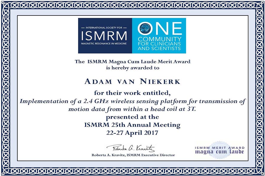
PhD student Adam van Niekerk and co-authors including Ernesta Meintjes were awarded an ISMRM Magna Cum Laude Merit Award and one of 200 power pitch presentations selected from 6700 abstracts at the recent ISMRM 25th Annual Meeting and Exhibition in Honolulu, Hawaii. Adam presented the implementation of a 2.4 GHz wireless sensing platform for transmission of motion data from within a head coil at 3T.
Please also see http://www.bme.uct.ac.za/bme/news-niekerk-ismrm2017award.
MRI research group abstracts to be presented at OHBM annual meeting
Abstracts by Martha Holmes, Emmanuel Nwosu, Jia Fan and Werner Stoltsz were accepted for presentation at the 2017 annual meeting of the Organization for Human Brain Mapping to be held in Vancouver from June 25-29. Congratulations to Werner, who was awarded an OHBM merit travel stipend to attend the meeting.
At the recent meeting of the Society for MR Radiographers & Technologists (SMRT) in Honolulu, Petty Samuels presented a poster titled Reproducibility of Brain Morphometry from MRI Scans Performed at Different Times of Day. Ingrid Op’t Hof presented pedriatric neuroimaging acquisition and analysis success rates at ages 5 and 7 years. They both received SMRT travel scholarships to attend the conference.
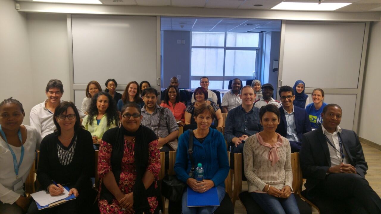
CUBIC UCT held its second Cardiovascular Magnetic Resonance workshop on Friday 31st March - Sunday 2nd April 2017 at Groote Schuur Hospital.
This three day hands-on Cardiovascular Magnetic Resonance Workshop is specifically designed to benefit Radiographers, Radiologists and Cardiologists working on, or interested in MRI, both in a clinical and research environment.
The workshop consisted of a series of lectures, followed by practical sessions based on the theory. During the hands-on sessions, participants had the opportunity to experiment with MRI where they gained a deeper understanding on how to plan, execute and analyse Cardiac MRI scans.
Mrs Petronella Samuels and colleagues from the division of cardiology recently presented the case study of a young woman with rheumatic heart disease at the annual meeting of the Society for Cardiovascular Magnetic Resonance. Investigating the cause of a conduction abnormality using multiparametric cardiovascular magnetic resonance imaging including cine, cine tagging, T2-weighted (STIR), pre contrast T1 and T2 mapping and late gadolinium enhancement (LGE) imaging, they were able to find evidence of myocarditis and valvulitis.
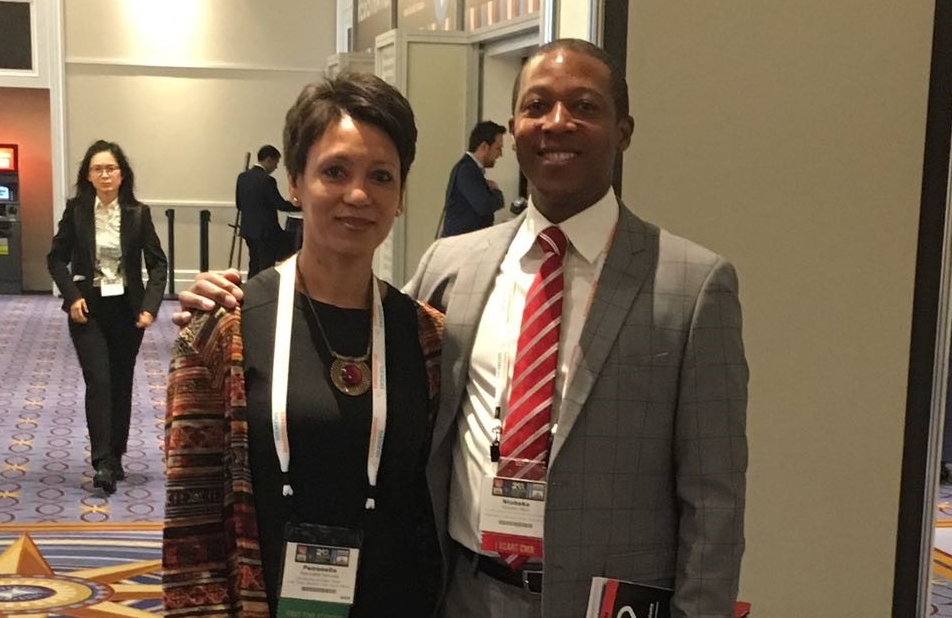
Image: Mrs Petty Samuels and Dr Ntobeko Ntusi